Port City Veterinary Referral Hospital
Introduction
This case study explores a complex medical scenario involving a canine patient referred to Port City Veterinary Referral Hospital due to a severe hemothorax caused by migrating porcupine quills. This document provides a detailed account of the patient's clinical presentation, diagnostic findings, surgical intervention, and post-operative care. Primary Care Team includes Surgeon: Dr. Robert Hillman, DVM, DACVS, Imaging: Dr. Mason Holland VMD, DACVR, and Anesthesiologist: Dr. Ella Pittman DVM, DACVAA
Patient Overview
Animal Name: Mouse Roy
Species: Canine
Sex: Male Age: 5.5 Years Breed: Mixed
Clinical Presentation
Mouse was initially treated for porcupine quilling three weeks prior to referral. Multiple quills were removed from the thoracic region at various times by different providers. Despite these efforts, the patient exhibited worsening respiratory distress, characterized by an increased respiratory rate and decreased appetite, culminating in bouts of vomiting and unsettled behavior at night.
Physical Examination Findings (05-28-2024):
General Appearance: Fearful, cautious handling required
Cardiovascular System: Normal rate and rhythm, no murmurs
Respiratory System: Slightly tachypneic with muffled breath sounds bilaterally
Gastrointestinal System: Non-painful abdomen, no masses or fluid wave detected
Musculoskeletal and Integumentary Systems: Ambulatory, no pain or lameness, no external quills observed
Neurological Exam: Normal with no deficits
Diagnostic Imaging
Dr. Mason Holland VMD, DACVR
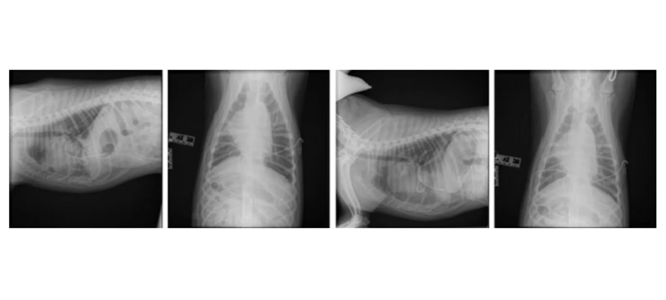
Radiographs (05-28-2024):
Findings: Mild residual pleural effusion, small amount of pleural air, presence of chest tubes
Interpretation: Post-chest tube placement with no primary intrathoracic lesions identified
Thoracic CT Scan (05-29-2024):
Findings: While the CT scan focused our attention on the cranial mediastinum, the quills themselves were not apparent on the imaging. This ambiguity necessitated a comprehensive approach to ensure that no lung area, left or right, was overlooked.
Interpretation: The CT findings were critical in guiding the decision for surgical exploration, leading to the choice of a median sternotomy to access the entire chest space for thorough examination and treatment.
Surgical Intervention
Procedure: Median sternotomy performed on 05-29-2024
Surgeon: Dr. Robert Hillman, DVM, DACVS,
Anesthesiologist: Ella Pittman, DVM, DACVAA
Surgical Findings:
A quill was located penetrating the right side of the cranial vena cava with additional quills in the pericardium.
A residual volume of frank blood was noted in the thoracic cavity, along with damage to the medial side of the right cranial lung lobe.
Procedure Details:
A ventral midline incision was made followed by an oscillating saw median sternotomy. Removal of the quill from the vena cava and exploratory thoracotomy were performed.
Closure utilized orthopedic wire and layered suturing techniques
Postoperative Care and Monitoring
Incision Care: Mouse’s incision was closed using intradermal sutures to minimize irritation and the potential for self-inflicted trauma. An Elizabethan collar was recommended to prevent licking or chewing at the incision site.
Monitoring for Complications: Close observation was required for signs of infection, increased redness, or discharge from the incision site. Regular follow-up appointments were scheduled to monitor healing.
Quilling Recommendations
Unfortunately, most dogs who get into trouble with porcupines do not learn from their mistakes.
The best defense against porcupine quills is prevention. The best way to prevent quilling is to advise pet owners to keep their dogs on a leash. Avoid allowing the dogs to roam at dusk or after dark and prevent them from going into areas with known porcupine dens.
Should Mouse get quilled again in the future, there are a few things the owner can do before reaching the vet.
Minimize movements and prevent him from rubbing his face if there are quills present. Manipulation of the quills may drive them deeper into the tissues, making it harder to retrieve them.
Do NOT cut the quills - cutting the shaft makes the quill splinter more easily which ultimately makes it harder to remove. It may also allow for segments of quills to become lodged in the tissues.
Do NOT attempt to remove quills yourself. Removing porcupine quills without the benefit of sedation or anesthesia and potent pain relief is extremely painful. This can result in a struggle, which can push the quills deeper, and a dog may lash out and bite, without meaning to hurt you.
Conclusion
This case highlights the challenges of managing hemothorax in canines resulting from foreign body migration, demonstrating the need for timely intervention and the complexities associated with extracting embedded quills from vital structures. Mouse’s case serves as an educational cornerstone for veterinary professionals handling similar emergency cases.
This document is intended for educational purposes to inform veterinary professionals about the potential complications and necessary interventions associated with porcupine quills in canines.


